
Share Important Moment of MileCell Bio with You
2025.09.14

Groundbreaking Research Insights
Emerging evidence shows that stellate cell lactate transporter MCT1 significantly promotes liver fibrosis by increasing type I collagen expression in vitro and in vivo, while MCT1 silencing attenuates TGF-β1-stimulated collagen production in human LX2 stellate cells[3].
Similarly, inhibiting nucleophosmin (NPM) – highly expressed in fibrotic livers and activated HSCs – reduces fibrotic markers while suppressing HSC proliferation and migration. Genetic or proteomic NPM inhibition dramatically mitigates carbon tetrachloride-induced fibrogenesis in murine models[4].
Your Gateway to Fibrosis Breakthroughs
Targeting hepatic stellate cells (HSCs) represents a promising therapeutic strategy. Milecell Bio delivers premium primary HSCs engineered for research excellence:
3 Game-Changing Advantages of Our HSCs
1. Physiologically Authentic Performance
Primary isolation preserves in vivo activation traits, enabling precise modeling of fibrotic progression.
2. Purity Perfection
Dual-gradient centrifugation + specific marker identification eliminates fibroblast contamination, ensuring data reliability.
3. Cryopreservation Revolution
-196°C LN₂ flash-freezing guarantees ready-to-use convenience – faster than instant noodles!
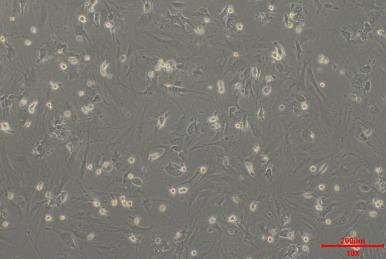
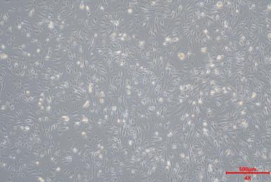
SD HSC -D1 BE HSC -D4 CY HSC -D1
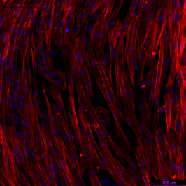
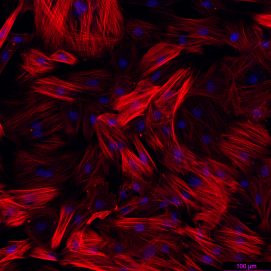
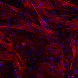
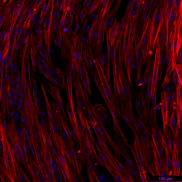
BE-HSC SD-HSC CY-HSC
Product Portfolio
| LOT | Product | Specification |
|---|---|---|
| CC57-HSC | Cryopreserved Mouse HSCs (Male, C57BL/6) | 0.5M |
| CCD-HSC | Cryopreserved Mouse HSCs (Male, CD-1) | 0.5M |
| CSD-HSC | Cryopreserved Rat HSCs (Male, SD) | 0.5M |
| CBD-HSC | Cryopreserved Canine HSCs (Male, Beagle) | 0.5M |
| CCY-HSC | Cryopreserved Monkey HSCs (Male, Cynomolgus) | 0.5M |
| CBM-HSC | Cryopreserved Minipig HSCs (Male, Bama) | 0.5M |
References
[1] Isaac, R. et al. (2024). Cell Metab 36(5):1030-1043. doi:10.1016/j.cmet.2024.04.003
[2] Aagaard, L. & Rossi, J.J. (2007). Adv Drug Deliv Rev 59(2-3):75-86. doi:10.1016/j.addr.2007.03.005
[3] Min, K. et al. (2024). eLife 12:RP89136. doi:10.7554/eLife.89136
[4] Ding, X. et al. (2023). Cell Death Dis 14(8):575. doi:10.1038/s41419-023-06043-0
