Share Important Moment of MileCell Bio with You
2025.04.11
Kupffer cells directly interact with bacterial components from the gut, including microbes, cell fragments, and endotoxins—which are considered substances that trigger macrophage activation. Once stimulated, these specialized macrophages secrete signaling molecules, such as cytokines, prostaglandins, nitric oxide, and oxidative free radicals. These mediators modulate the functional characteristics of Kupffer cells themselves and neighboring cell populations, such as hepatocytes, stellate cells, vascular endothelial cells, and transient immune cells traversing the liver tissue. Kupffer cells play a key role in coordinating the liver's adaptation to pathological damage (including infections, toxic substances, ischemic injury, surgical resection, and metabolic disorders).
Due to their multifunctionality, Kupffer cells are an important component in in vitro applications, such as drug screening programs, toxicity assessment, modeling of inflammation-related drug toxicity, and simulation of disease mechanisms. Scientists use these cells in various experimental models, ranging from isolated monoculture to multicellular co-culture systems and engineered liver microtissue platforms.
Kupffer Cells from MileCell Bio
MileCell Bio’s Animal Hepatic Kupffer cells are isolated from rat, mouse, dog, monkey liver tissue. Each vial contains 0.5 million cells in 1 mL volume.
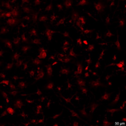
1. Immunofluorescence Staining of CD68

SD Kupffer Cells
2. Photomicrograph

SD Kupffer -D1 CD-1 Kupffer -D2
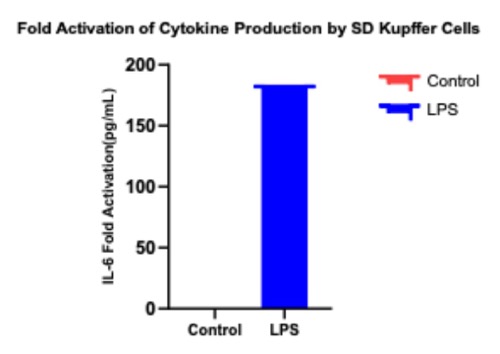
3. LPS Stimulation

Fold activation of cytokine production by Kupffer cells. Cryopreserved SD Rat Kupffer cells were thawed and plated in 48-well plates in Kupffer Culture Medium. After 1 day in culture and without any medium replacement, cells were treated with 1 ug/ml LPS for 24 hours. At the end of the treatment period, RNA was isolated for mRNA analysis. Results were expressed in terms of fold activation of IL-6.
